ResearchLayout & EquipmentAlumniStudentsDonorsTeamMulticentre & Markerless Orthopedic Assessment WikiContact
The impact of biomechanics research has historically been limited by the difficulties associated with marker-based motion capture systems, which require skilled operators and significant time for data collections. Recent advances in computer vision technology have led to the proliferation and improvement of video-based human motion tracking systems. Our lab has partnered with Theia Markerless, Inc., a Kingston-based company on the leading edge of markerless motion capture technology to test, validate, and utilize their markerless motion capture software Theia3Din biomechanics research contexts.
Our partnership with Theia Markerless has led to three peer-reviewed publications to date [1, 2, 3], which have laid the groundwork for several exciting upcoming studies on the implementation of Theia3D markerless motion capture in wider contexts, such as in clinical and outdoor settings. We are excited to be initiating research projects that go beyond what was previously possible with marker-based motion capture, including large-scale outdoor data collections and building movement datasets of populations with gait-affecting pathologies and orthopaedic surgery patients.
Our long-term goal is to develop effective tools for the assessment and treatment of musculoskeletal disorders, and help people maintain active lives longer.
The state-of-the-art facility offers a unique means to measure the mechanical factors of joint loading, orientation, and neuromuscular function during activities of daily living including high demand recreational and occupational tasks.
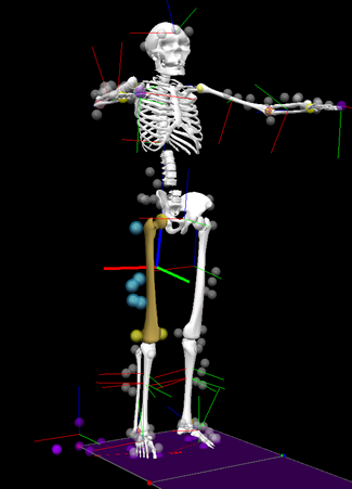
The core technology for this analysis is human motion capture. Our lab features traditional marker-based motion capture, which uses optoelectronic infrared cameras to the measure the three-dimensional position of retroreflective spherical markers fastened to palpable bony landmarks, as well as markerless motion capture, which uses machine vision and deep learning algorithms to estimate skeletal landmark positions directly.
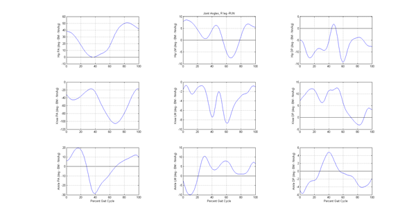
By tracking several landmarks on each body segment, the full pose (position and orientation) of each segment can be estimated throughout the performance of dynamic tasks, enabling measures such as joint positions and joint angles to be obtained.

Force platforms embedded in the ground are used to measure the magnitude, direction, and location of the ground reaction force during dynamic tasks.
Combining the ground reaction forces, with the limb segment pose data provides the basis for calculating joint loading (net reaction moments and forces).

To extend testing to high performance activity, the lab also includes a secondary marker-based motion capture system surrounding an instrumented treadmill. This technology is capable of measuring ground reaction forces over multiple gait cycles and even during uphill or downhill inclines. An instrumented stairway allows collection of motion and force data during stair climb and decent.
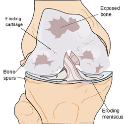
Osteoarthritis is a common age-related impairment that can cause pain and physical disability. The factors that influence the initiation and progression of OA are not well understood. There is a lack of data on the pathomechanics of this disease as well as causes for this disease to progress more rapidly in some individuals than others.
Biomechanics studies have demonstrated the profound changes associated with severe OA, including differences in stride characteristics, joint kinematics, kinetics and neuromuscular function. More recent studies of moderate knee OA reveal the importance of joint kinetics in understanding the pathomechanics of OA and in the design and evaluation of treatment options.
Non-surgical treatments aimed at improving the mechanical environment of the knee such as bracing, heel wedges, gait modifications and therapeutic techniques have shown promise in the management of the symptoms of OA.
